Click image to see more details
-

-
-
-
-
+2
Product Info Summary
| SKU: | A03154-1 |
|---|---|
| Size: | 100 μg/vial |
| Reactive Species: | Human, Mouse, Rat |
| Host: | Rabbit |
| Application: | Flow Cytometry, IF, IHC, ICC, WB |
Customers Who Bought This Also Bought

Product info
Product Name
Anti-Synaptopodin/SYNPO Antibody Picoband™
View all Synaptopodin Antibodies
SKU/Catalog Number
A03154-1
Size
100 μg/vial
Form
Lyophilized
Description
Boster Bio Anti-Synaptopodin/SYNPO Antibody Picoband™ catalog # A03154-1. Tested in Flow Cytometry, IF, IHC, ICC, WB applications. This antibody reacts with Human, Mouse, Rat.
Storage & Handling
Store at -20˚C for one year from date of receipt. After reconstitution, at 4˚C for one month. It can also be aliquotted and stored frozen at -20˚C for six months. Avoid repeated freeze-thaw cycles.
Cite This Product
Anti-Synaptopodin/SYNPO Antibody Picoband™ (Boster Biological Technology, Pleasanton CA, USA, Catalog # A03154-1)
Host
Rabbit
Contents
Each vial contains 4mg Trehalose, 0.9mg NaCl, 0.2mg Na2HPO4, 0.01mg NaN3.
Clonality
Polyclonal
Isotype
Rabbit IgG
Immunogen
A synthetic peptide corresponding to a sequence at the C-terminus of human Synaptopodin/SYNPO, which shares 100% and 94.3% amino acid (aa) sequence identity with mouse and rat Synaptopodin/SYNPO, respectively.
*Blocking peptide can be purchased. Costs vary based on immunogen length. Contact us for pricing.
Cross-reactivity
No cross-reactivity with other proteins.
Reactive Species
A03154-1 is reactive to SYNPO in Human, Mouse, Rat
Applications
A03154-1 is guaranteed for Flow Cytometry, IF, IHC, ICC, WB Boster Guarantee
Observed Molecular Weight
99 kDa
Calculated molecular weight
99.463kDa
Background of Synaptopodin
The spine apparatus (SA) is a specialized form of endoplasmic reticulum (ER) that is found in a subpopulation of dendritic spines in central neurons. The SA consists of a series of stacked discs that are though to be connected to each other and to the dendritic system of ER-tubules. The actin binding protein synaptopodin (which has originally been described in podocytes of the kidney) is an essential component of the SA. Mice that lack the gene for synaptopodin do not form a spine apparatus. The SA is believed to play a critical role in learning and memory. In summary, an important function of the spine apparatus is the regulation of plasticity at individual synapses, a process known as metaplasticity. The International Radiation Hybrid Mapping Consortium mapped the SYNPO gene to chromosome 5.
Antibody Validation
Boster validates all antibodies on WB, IHC, ICC, Immunofluorescence, and ELISA with known positive control and negative samples to ensure specificity and high affinity, including thorough antibody incubations.
Innovating Scientists Reward
If you are the first to review this product, or if you have results for a special sample, species or application this product is not validated in, share your results with us and receive product credits you can use towards any Boster products! Applicable to all scientists worldwide.
Submit A Review
Assay dilution & Images
Reconsitution
Add 0.2ml of distilled water will yield a concentration of 500ug/ml.
Assay Dilutions Recommendation
The recommendations below provide a starting point for assay optimization. The actual working concentration varies and should be decided by the user.
Western blot, 0.1-0.25μg/ml, Human, Mouse, Rat
Immunohistochemistry (Paraffin-embedded Section), 2-5μg/ml, Human, Mouse, Rat
Immunocytochemistry/Immunofluorescence, 5μg/ml, Human
Flow Cytometry, 1-3μg/1x106 cells, Human
Validation Images & Assay Conditions
Click image to see more details
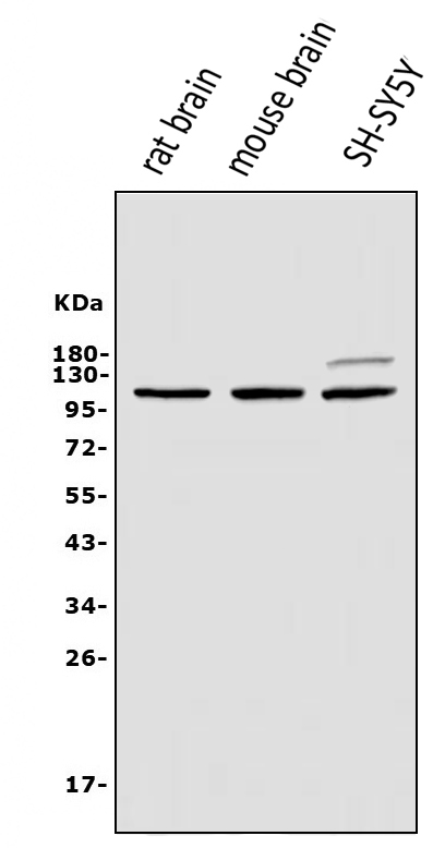
Figure 1. Western blot analysis of Synaptopodin/SYNPO using anti-Synaptopodin/SYNPO antibody (A03154-1).
Electrophoresis was performed on a 5-20% SDS-PAGE gel at 70V (Stacking gel) / 90V (Resolving gel) for 2-3 hours. The sample well of each lane was loaded with 50ug of sample under reducing conditions.
Lane 1: rat brain tissue lysates,
Lane 2: mouse brain tissue lysates,
Lane 3: human SH-SY5Y whole cell lysates.
After Electrophoresis, proteins were transferred to a Nitrocellulose membrane at 150mA for 50-90 minutes. Blocked the membrane with 5% Non-fat Milk/ TBS for 1.5 hour at RT. The membrane was incubated with rabbit anti-Synaptopodin/SYNPO antigen affinity purified polyclonal antibody (Catalog # A03154-1) at 0.25 μg/mL overnight at 4°C, then washed with TBS-0.1%Tween 3 times with 5 minutes each and probed with a goat anti-rabbit IgG-HRP secondary antibody at a dilution of 1:5000 for 1.5 hour at RT. The signal is developed using an Enhanced Chemiluminescent detection (ECL) kit (Catalog # EK1002) with Tanon 5200 system. A specific band was detected for Synaptopodin/SYNPO at approximately 99KD. The expected band size for Synaptopodin/SYNPO is at 99KD.

Click image to see more details
Figure 2. IHC analysis of Synaptopodin/SYNPO using anti-Synaptopodin/SYNPO antibody (A03154-1).
Synaptopodin/SYNPO was detected in paraffin-embedded section of rat brain tissue. Heat mediated antigen retrieval was performed in EDTA buffer (pH8.0, epitope retrieval solution). The tissue section was blocked with 10% goat serum. The tissue section was then incubated with 2μg/ml rabbit anti-Synaptopodin/SYNPO Antibody (A03154-1) overnight at 4°C. Biotinylated goat anti-rabbit IgG was used as secondary antibody and incubated for 30 minutes at 37°C. The tissue section was developed using Strepavidin-Biotin-Complex (SABC) (Catalog # SA1022) with DAB as the chromogen.

Click image to see more details
Figure 3. IHC analysis of Synaptopodin/SYNPO using anti-Synaptopodin/SYNPO antibody (A03154-1).
Synaptopodin/SYNPO was detected in paraffin-embedded section of human renal cancer tissue. Heat mediated antigen retrieval was performed in EDTA buffer (pH8.0, epitope retrieval solution). The tissue section was blocked with 10% goat serum. The tissue section was then incubated with 2μg/ml rabbit anti-Synaptopodin/SYNPO Antibody (A03154-1) overnight at 4°C. Biotinylated goat anti-rabbit IgG was used as secondary antibody and incubated for 30 minutes at 37°C. The tissue section was developed using Strepavidin-Biotin-Complex (SABC) (Catalog # SA1022) with DAB as the chromogen.

Click image to see more details
Figure 4. IF analysis of Synaptopodin/SYNPO using anti-Synaptopodin/SYNPO antibody (A03154-1).
Synaptopodin/SYNPO was detected in immunocytochemical section of U20S cells. Enzyme antigen retrieval was performed using IHC enzyme antigen retrieval reagent (AR0022) for 15 mins. The cells were blocked with 10% goat serum. And then incubated with 5μg/mL rabbit anti-Synaptopodin/SYNPO Antibody (A03154-1) overnight at 4°C. DyLight®594 Conjugated Goat Anti-Rabbit IgG (BA1142) was used as secondary antibody at 1:100 dilution and incubated for 30 minutes at 37°C. The section was counterstained with DAPI. Visualize using a fluorescence microscope and filter sets appropriate for the label used.

Click image to see more details
Figure 5. Flow Cytometry analysis of U87 cells using anti-Synaptopodin/SYNPO antibody (A03154-1).
Overlay histogram showing U87 cells stained with A03154-1 (Blue line).The cells were blocked with 10% normal goat serum. And then incubated with rabbit anti-Synaptopodin/SYNPO Antibody (A03154-1, 1μg/1x106 cells) for 30 min at 20°C. DyLight®488 conjugated goat anti-rabbit IgG (BA1127, 5-10μg/1x106 cells) was used as secondary antibody for 30 minutes at 20°C. Isotype control antibody (Green line) was rabbit IgG (1μg/1x106) used under the same conditions. Unlabelled sample (Red line) was also used as a control.

Click image to see more details
Figure 6. IHC analysis of Synaptopodin/SYNPO using anti-Synaptopodin/SYNPO antibody (A03154-1).
Synaptopodin/SYNPO was detected in paraffin-embedded section of mouse brain tissue. Heat mediated antigen retrieval was performed in EDTA buffer (pH8.0, epitope retrieval solution). The tissue section was blocked with 10% goat serum. The tissue section was then incubated with 2μg/ml rabbit anti-Synaptopodin/SYNPO Antibody (A03154-1) overnight at 4°C. Biotinylated goat anti-rabbit IgG was used as secondary antibody and incubated for 30 minutes at 37°C. The tissue section was developed using Strepavidin-Biotin-Complex (SABC) (Catalog # SA1022) with DAB as the chromogen.
Protein Target Info & Infographic
Gene/Protein Information For SYNPO (Source: Uniprot.org, NCBI)
Gene Name
SYNPO
Full Name
Synaptopodin
Weight
99.463kDa
Superfamily
synaptopodin family
Alternative Names
KIAA1029; Synaptopodin; SYNPO SYNPO synaptopodin synaptopodin
*If product is indicated to react with multiple species, protein info is based on the gene entry specified above in "Species".For more info on SYNPO, check out the SYNPO Infographic

We have 30,000+ of these available, one for each gene! Check them out.
In this infographic, you will see the following information for SYNPO: database IDs, superfamily, protein function, synonyms, molecular weight, chromosomal locations, tissues of expression, subcellular locations, post-translational modifications, and related diseases, research areas & pathways. If you want to see more information included, or would like to contribute to it and be acknowledged, please contact [email protected].
Specific Publications For Anti-Synaptopodin/SYNPO Antibody Picoband™ (A03154-1)
Hello CJ!
No publications found for A03154-1
*Do you have publications using this product? Share with us and receive a reward. Ask us for more details.
Recommended Resources
Here are featured tools and databases that you might find useful.
- Boster's Pathways Library
- Protein Databases
- Bioscience Research Protocol Resources
- Data Processing & Analysis Software
- Photo Editing Software
- Scientific Literature Resources
- Research Paper Management Tools
- Molecular Biology Software
- Primer Design Tools
- Bioinformatics Tools
- Phylogenetic Tree Analysis
Customer Reviews
Have you used Anti-Synaptopodin/SYNPO Antibody Picoband™?
Submit a review and receive an Amazon gift card.
- $30 for a review with an image
Be the first to review Anti-Synaptopodin/SYNPO Antibody Picoband™
*The first user to submit a review for a product is eligible for Boster's Innovating Scientists Reward, which gives product credits. This is in addition to the gift card reward.
Customer Q&As
Have a question?
Find answers in Q&As, reviews.
Can't find your answer?
Submit your question
1 Customer Q&As for Anti-Synaptopodin/SYNPO Antibody Picoband™
Question
What tissue is tested in image 3 of the product page for A03154-1?
Verified customer
Asked: 2021-01-06
Answer
For the Anti-Synaptopodin/SYNPO Antibody Picoband™ (A03154-1) product page, it is human renal cancer tissue that is tested in image 3.
Boster Scientific Support
Answered: 2021-01-06
